page 126
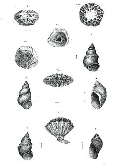
On a new genus of POLYZOA. P1. 13, fig., 1, 1a, 1b, 1c.
By the REV. J. E. TENISON-WOODS, F.L.S., &c.
The very interesting and curious genus which I now describe was dredged by the Hon. Wm. Macleay, F.L.S., off Darnley Island, at a depth of 10 or 20 fathoms, on coral mud. It belongs to the Cheilostomatous sub-order, but differs so completely from any of the described families that its affinities and relations must remain problematical until others are discovered, as no doubt in time there will be. The nearest family is the Selenariadoe, which has the polyzoary more or less orbicular, convex on one side, but there is no special modification of any organ in the manner seen in the species under consideration. Its singular beauty, both as regards design and ornament, renders it a remarkable addition to an order where beauty and variety are the rule. I shall distinguish the genus by the name Euktimenaria, from [greek], well built.
EUKTIMENARIA, new genus.
Polyzoary free, upper surface convex, covered with cells; lower surface divided into five portions, each containing large pores; in the centre of the base a vermiculate quinque-partite body.
EUKTIMENARIA DUCALIS. Pl. 13, fig. 1, 1a, 1b, 1c.
Convex, with pentagonal outline; the edge circumscribed by a raised margin of five arches, whence it descends to a broad pentagonal pedicel by five arched concave surfaces, which are horizontally divided in the centre by a straight raised double ridge, above and below the centre of which there is a large conspicuous pore; the pore above is semi-circular, that below is
page 127
perfectly round; both seem deep. The margin of each of the arched spaces curves round into a loop at each side below the lower pore, and is curved again in a contrary direction at each side so as to form another small loop in which there is another small pore. Beneath the lower of the two large central pores there are one or two conspicuous grooves to the base. Upper convex surface covered with concave cells, with a distinct raised margin; mouth in the centre, semi-circular; with a raised margin. Shape of cells from oval to circular, a few almost pentagonal; the centre of the convex surface seems covered with cells, but they are worn almost smooth on both the specimens. The base is vermiculate, but with a radiate tendency, and forming a quinque-partite pattern. Between the margin of the five sides there are upper and lower angular spaces, giving great elegance to the design.
Dimensions: Alt. 6, diam. of summit 8, of base 4 1/2, lat. of 5 lateral spaces 4 1/2, alt. 3 1/2 millim.
I am unable to suggest any explanation of the pores on the sides or the organs which form the margins, transverse bands, &c. It is quite evident that there must be some individuality in these zoothomes, apart from what we call the animal which dwells in the cells, or the symmetrical arrangements in this specimen could not be explained. Only two were found by the Chevert Expedition in all their dredgings, and both these were a little worn as if they had been dead some time. There is something in the species which recalls the elegent forms of Polyzoa in the European chalk, but there is no fossil form that I have heard of which nearly resembles it. There are fossils from the Maestricht Chalk which seem to have some analogy with Euktiminaria (one species is named Glenotremites paradoxus by Goldfuss), and geologists are not agreed upon their position in classification or their character. M. d'Orbigny considered them as Comatulae without their arms, but there were reasons for rejecting this view. The mouth (?) was surrounded by five funnel-shaped openings and five petaloid grooves which were probably places for the insertion of arms, All the surface
page 128
of the calice was surrounded by perforated depressions which, according to Agassiz, may have been articulations for dorsal rays. There is another species from the chalk of Rugen with the 5 funnel-shaped openings reduced to little pores.
EXPLANATION OF PLATE 13.
Fig. 1. Euktiminaria ducalis, side view, slightly enlarged.
Fig. 1a. ditto ditto seen from above.
Fig. 1b. Mouth of cell much enlarged.
Fig. 1c. Base slightly enlarged.